CARDIOVASCULAR MAGNETIC RESONANCE IMAGING (CMR)
CONTRIBUTED BY: DR. DUANE CHAMBERS – CONSULTANT RADIOLOGIST
Cardiovascular disease remains the leading cause of death in Jamaica. Since 2023, Northcoast Imaging
(NCI) has proudly offered advanced cardiac MRI services in the Western region, helping to bridge the
diagnostic gap in cardiac care across Jamaica and the wider Caribbean.
Cardiovascular magnetic resonance imaging (CMR) is now widely regarded as the gold standard for
evaluating myocardial function, tissue characterization, and structural heart disease. It plays a critical role in
the evaluation of conditions such as congenital heart disease, cardiomyopathies, myocarditis, pericardial
disease, and coronary artery disease.
LEADING INDICATIONS FOR CARDIAC MRI AT NCI
- Coronary artery disease
- Congenital heart disease
- Acquired diseases of the vasculature
- Cardiomyopathies
- Pericardial disease
- Cardiac masses
- Valvular heart disease
- Myocardial viability testing
MYOCARDIAL VIABILITY TESTING
At NCI, we utilize high-quality CMR to define infarct size, assess tissue at risk, and predict myocardial
recovery potential. Using techniques such as late gadolinium enhancement, stress perfusion imaging, and
tissue mapping, we provide detailed, non-invasive insights to guide patient management, particularly in cases
of ischemic heart disease.
ADVANTAGES OF CARDIAC MRI
- Superior image quality and diagnostic clarity
- No ionising radiation
- Precise ejection fraction assessment
- Tissue characterization (e.g., edema, fibrosis, scar)
- Effective in evaluating chest pain, especially post-COVID or post-vaccination myocarditis
- Essential in pre-surgical planning (e.g., bypass surgery, valve repair)
LIMITATIONS TO CONSIDER
- Not suitable for patients with certain implants or retained metal fragments
- May be difficult for patients with severe claustrophobia
- Gadolinium contrast is contraindicated in advanced renal disease
- MRI scanner size and weight limits may restrict some patients
CMR IN THE POST-COVID ERA
Myocarditis continues to be a relevant diagnosis in patients with unexplained chest pain, especially following
viral illness or vaccination. At NCI, we use updated Lake Louise criteria and advanced parametric mapping
techniques to diagnose these cases accurately and guide long-term management.
CLOSING NOTE
Since the launch of cardiac MRI services in 2023, NCI has proudly expanded its capabilities to meet the
growing demand for precision cardiac imaging. With a commitment to cutting-edge technology and expert
analysis, we are helping clinicians across the region make clearer, faster, and more confident cardiovascular
diagnoses.
Questions or referrals?
info@northcoastimaging.com
876-684-5656 | 876-619-1027 | 876-579-9533 (Digicel)
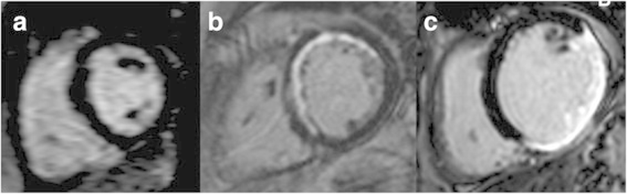
LGE on CMR in 3 different patients. a- No scar, b- Less than 50 % thickness involvement in multiple segments and c- More than 50 % thickness involvement in multiple segments
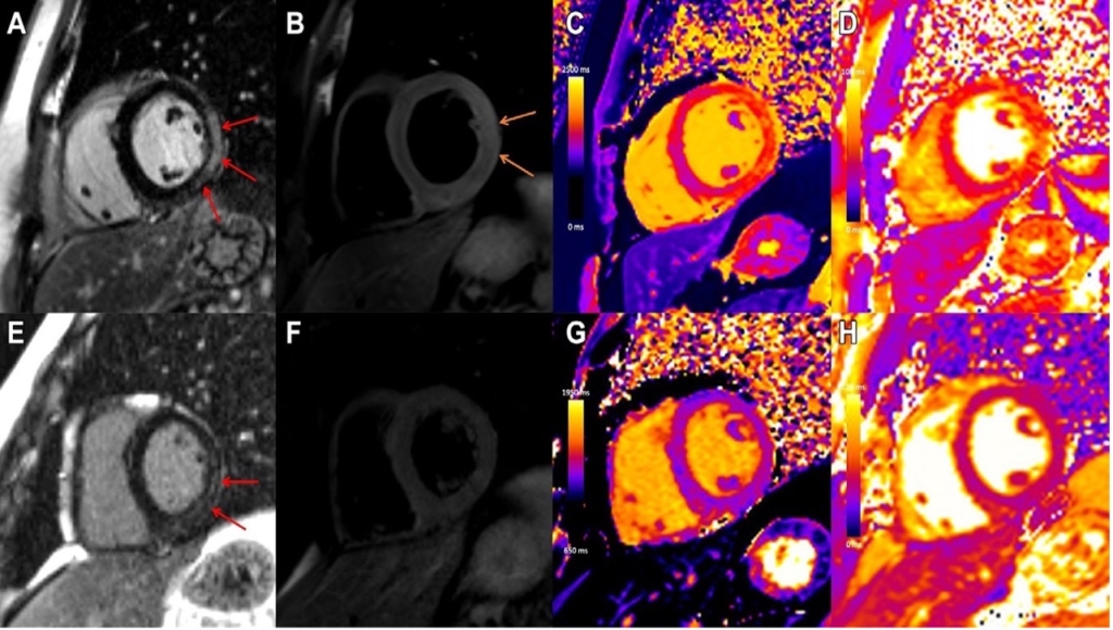
Case example in a 31-year-old man with viral myocarditis. Initial cardiac MRI performed at 3 T within 1 week of symptom onset demonstrates (A) subepicardial to nearly transmural late gadolinium enhancement (LGE) at the basal to mid anterior lateral, inferior lateral, and inferior wall (red arrows) with (B) corresponding high T2 signal, in keeping with oedema (orange arrows), (C) high regional native T1 (1480 msec), and (D) high regional native T2 (56 msec) on short-axis images. Images from follow-up cardiac MRI performed at 1.5 T 5 months later demonstrate contraction of subepicardial LGE at the basal to mid inferior lateral and inferior wall (E, red arrows) with (G) corresponding high regional native T1 suggestive of fibrosis (1305 msec) and (F) resolution of oedema with no corresponding high T2 signal and (H) normalization of T2 mapping values (46 msec)

T2* < 20 ms suggests iron overload. T1 also decreases